Description
Overview
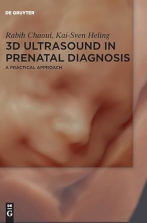
In the last decade, 3D ultrasound has become widely used in obstetrical imaging. It is estimated that more than half of obstetrical clinics now use ultrasound equipment with 3D capabilities. Initially recognized for producing beautiful images of fetal faces, 3D ultrasound has evolved into an essential tool for prenatal diagnosis due to its ability to visualize fetal organs in both normal and abnormal conditions. This book serves as a state-of-the-art, practical guide to the application of 3D ultrasound in obstetrics, offering valuable insights for clinical practice.
Key Features
- Comprehensive coverage of 3D ultrasound technology and its role in prenatal diagnosis.
- A structured approach to the technical principles of 3D ultrasound.
- Step-by-step explanations of various 3D rendering tools and their applications.
- Detailed exploration of the clinical use of 3D ultrasound in examining fetal organs.
- Richly illustrated with high-quality images demonstrating the clinical utility of 3D ultrasound.
Highlights
This book provides a practical, evidence-based resource for integrating 3D ultrasound into obstetrical practice. It includes step-by-step instructions for using 3D rendering tools and detailed discussions on their applications in diagnosing fetal conditions. The book is designed to enhance clinical confidence, offering key takeaways and summaries to reinforce learning.
Book Structure
The book is divided into three sections:
- Technical Principles – Covers the fundamental concepts behind 3D ultrasound technology.
- 3D Rendering Tools – Provides a step-by-step guide to different 3D imaging techniques and their applications.
- Clinical Applications – Focuses on the use of 3D ultrasound in fetal organ examination and prenatal diagnosis.
Authors
Rabih Chaoui, Kai-Sven Heling
Book Preview:

